LaMore, T. BS, BA, et al.
Abstract:
(Final manuscript in progress)
Background
The Locus coeruleus(LC) is one of the first sites of tau aggregation and suffers from dramatic volume loss early on. Due to the shrinking of the LC preceding the onset of clinical symptoms of AD by decades, a precise understanding of the neurobiological correlates of MRI signal may turn LC neuroimaging into an early non-invasive staging biomarker for AD. Though there are many efforts to image the LC in-vivo, first, it is vital to confirm postmortem validation of the MRI signal. In this study, we are attempting to validate postmortem histological reconstructions of the LC to its corresponding postmortem MRI reconstructions, voxel-to-voxel.
Method
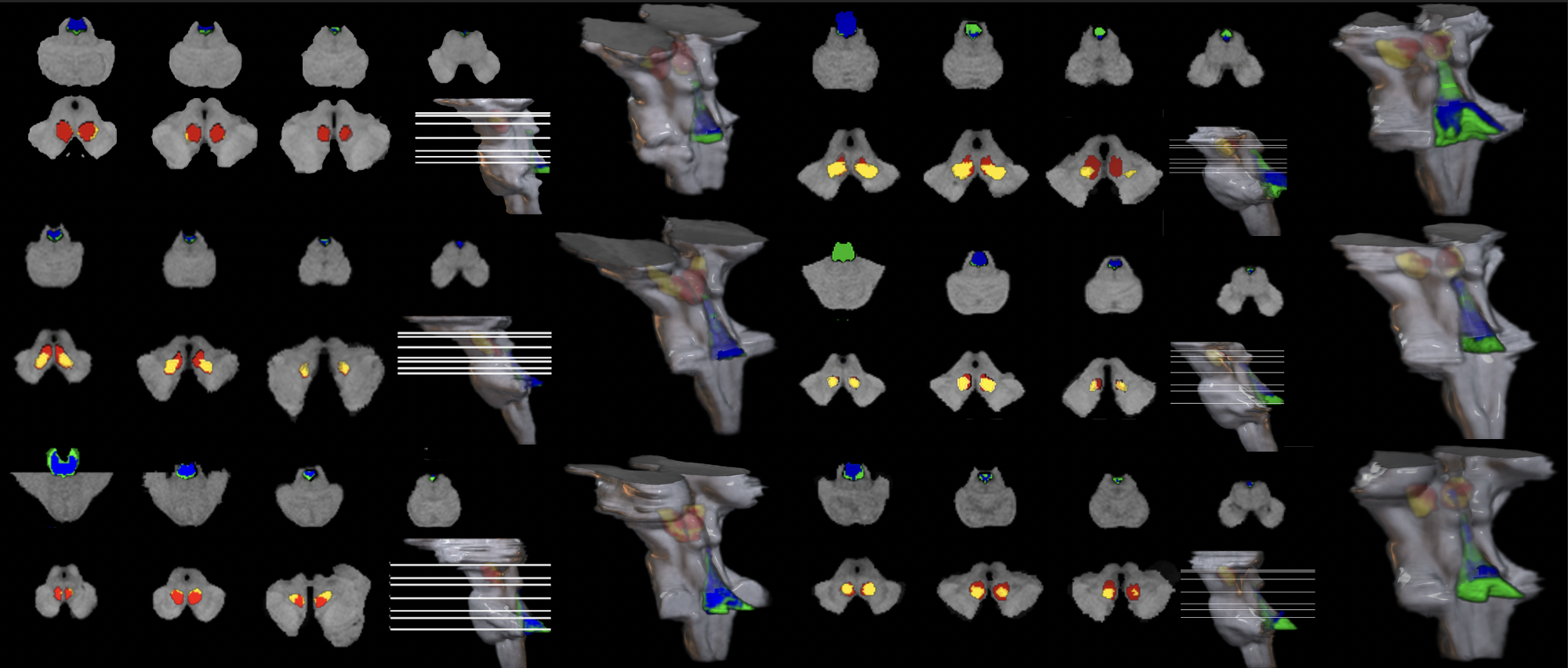
We used a pipeline developed in-house employing brainstem processing, staining, and computer-based algorithms for 2D and 3D registration to reconstruct the histological volume of the brainstem with structural detail (Fig 1). This pipeline allows for precise alignment of histology-based cytoarchitectural 3D reconstructions of the brainstem to its 7T-MRI counterpart. Photoshop and Freeview were used to manually segment the LC in digital images of cross sections of the brainstem labeled with Nissl staining, to generate 2D LC masks in all 6 cases. We then registered the stained cross sections and their corresponding LC masks to the cross sections’ blockface to correct for any deformation of tissue during the staining process. We applied Freeview commands to reconstruct the 3D brainstems and their 3D LC masks. We registered the resulting 3D reconstruction and LC mask to matching MRI coordinates, enabling voxel-to-voxel comparisons. We will continue to demonstrate the value of our pipeline in regards to validating neuroimaging methods in AD with these structural masks of the postmortem LC registered to the MRI counterpart in all 6 cases.
Result
The histological 3D LC masks were registered onto the parallel 3D MRIs. In Freeview, we plan to run correlation analysis with an output of dice coefficient ranging from 0-1, with a coefficient of 1 translating to the voxels of the LC masks and the MRI signal overlapping exactly.
Conclusion
Our pipeline enables voxel-to-voxel correlation analysis between histology and MRI scans for validation of LC neuroimaging. Current finalization of 3 other cases is ongoing.


